
$

PAUS ID

Padjadjaran Authentication System.

$

Portal Mahasiswa

Merupakan portal informasi bagi mahasiwa UNPAD

$

Portal Staff

Merupakan portal informasi bagi staf UNPAD.

$

Email UNPAD

Layanan email berbasis Google App for Education (GAFE) bagi seluruh sivitas akademika Unpad.

UNPAD

Histopathological Progression and Clinicopathologic Factors Associated with Anogenital Condyloma Acuminata: A Pathological Perspective From a Tertiary Hospital in Indonesia

Histopathological Progression and Clinicopathologic Factors Associated with Anogenital Condyloma Acuminata: A Pathological Perspective From a Tertiary Hospital in Indonesia

44

Authors

Karimah Ihda Husnayain1, Hermin Aminah Usman1,Etis Primastari1, Pati AjiAchdiat2

Affiliations

1Department of Anatomic Pathology, Faculty of Medicine, Universitas Padjadjaran, Dr. Hasan Sadikin

Hospital, Bandung, West Java, Indonesia 2Department of Dermatology and Venereology, Faculty of Medicine, Universitas Padjadjaran, Dr. Hasan Sadikin Hospital, Bandung, West Java, Indonesia

Background

• Anogenital condyloma acuminata (CA) is a benign lesion caused by low-risk HPV.

• A subset of lesions can progress histopathologically, and HIV-related immunosuppression may change their course.

Methods

• Retrospective cross-sectional study of 224 patients with histopathologically confirmed CA at Dr. Hasan Sadikin General Hospital, Bandung (2023–2024).

• Clinical, pathological, and sexual behavior data analysed with chi-square/Fisher tests and logistic regression.

Key Results

• 87 patients (38.8%) were HIV-positive.

• Bisexual orientation, lesion duration over 6 months, and lesion location were independent predictors of HIV positivity.

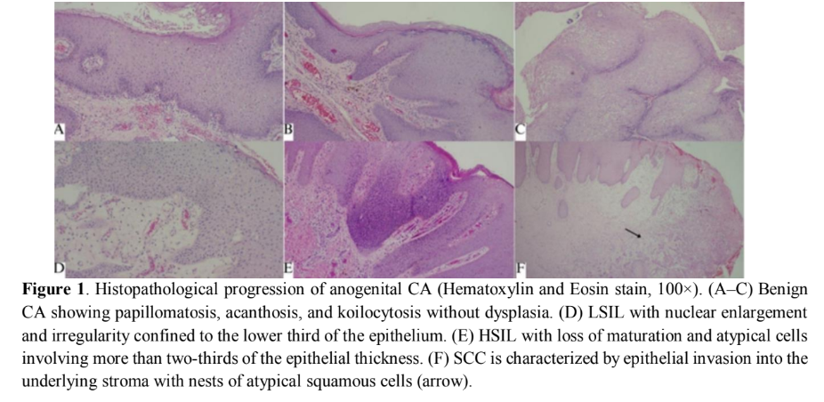
• Histopathological progression occurred in 13 patients (5.8%), including two squamous cell carcinomas.

Discussion & Conclusion

• HIV is highly prevalent among patients with CA.

• Routine histopathological evaluation is essential to detect malignant potential early.

Related SDGs

SDG 3: Good Health and Well-Being – Improving early detection and care for HPV- and HIV-related disease

Journal Information

Journal: Berkala Ilmu Kesehatan Kulit Dan Kelamin

DOI: https://doi.org/10.20473/bikkk.V38.1.2026.27-38

Exploring Trends in Endoscopic Dacryocystorhinostomy Research in Asia: A Bibliometric Analysis

Exploring Trends in Endoscopic Dacryocystorhinostomy Research in Asia: A Bibliometric Analysis

43

Authors

Josiah Irma 1,2,, Saraswati Anindita Rizki 3, Arief S. Kartasasmita 1,4, Angga Kartiwa 4 and Irawati Irfani 4

Affiliations

1Doctoral Program in Medical Sciences, Faculty of Medicine, Universitas Padjadjaran, Bandung 40161, Indonesia;

2Ophthalmology Department, Universitas Pelita Harapan, Tangerang 15810, Indonesia

3Faculty of Medicine, Universitas Pelita Harapan, Tangerang 15810, Indonesia;

4Ophthalmology Department, Faculty of Medicine, Universitas Padjadjaran, Bandung 40117, Indonesia;

Background

• Research trends in endoscopic dacryocystorhinostomy (Endo-DCR) across Asian countries had not been systematically mapped.

Methods

• Bibliometric analysis of the Scopus database, performed in April 2024.

• Data analysed with Microsoft Excel and VOSviewer.

Key Results

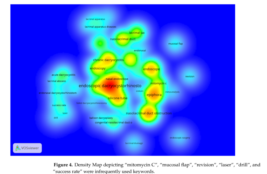
• 730 articles were included; publications rose from the early 2000s, with a surge after 2010.

• India and China were the leading contributors in Asia.

• Common keywords included endoscopy, silicone tube, and epiphora; mitomycin C, mucosal flap, and success rate were rarely used.

Discussion & Conclusion

• Endo-DCR publications grew markedly between 1972 and 2023, mostly from India and China.

• Topics such as mitomycin C, mucosal flap, laser, and success rate remain under-researched, pointing to future research opportunities.

Related SDGs

SDG 3: Good Health and Well-Being – Mapping evidence to improve surgical eye and ENT care

SDG 9: Industry, Innovation, and Infrastructure – Strengthening research and innovation through bibliometric insight

Journal Information

Journal: Surgeries

DOI: https://doi.org/10.3390/surgeries7010032

SOX2 expression in the primary tumor of castration-naive metastatic prostate adenocarcinoma in association with metastasis extent

SOX2 expression in the primary tumor of castration-naive metastatic prostate adenocarcinoma in association with metastasis extent

42

Authors
Meilania Saraswati¹, Aria Kekalih², Lisnawati³, Nur Rahadiani³, Asmarinah⁴, Bethy Suryawathy Hernowo⁵, Agus Rizal Ardy Hariandy Hamid⁶, Chaidir Arif Mochtar⁶

Affiliations

1Doctoral Program in Medical Sciences, Faculty of Medicine, Universitas Indonesia, Jakarta, Indonesia, 2Department of Community Medicine, Faculty of Medicine, Universitas Indonesia, Jakarta, Indonesia, 3Department of Anatomical Pathology, Faculty of Medicine, Universitas Indonesia, Cipto Mangunkusumo Hospital, Jakarta, Indonesia, 4Department of Medical Biology, Faculty of Medicine, Universitas Indonesia, Jakarta, Indonesia, 5Department of Anatomical Pathology Faculty of Medicine, Universitas Padjadjaran, Hasan Sadikin Hospital, Bandung, Indonesia, 6Department of Urology, Faculty of Medicine, Universitas Indonesia, Cipto Mangunkusumo Hospital, Jakarta, Indonesia

Background

• Poor prognosis in metastatic prostate adenocarcinoma may relate to stem cell-related genes.

• This study examined cancer stem cell markers in relation to metastasis extent.

Methods

• Cross-sectional analytical study of prostate specimens from Cipto Mangunkusumo Hospital.

• 61 patients aged 50+ (2020–2023) grouped as high-volume disease (n = 38) or low-volume disease (n = 23).

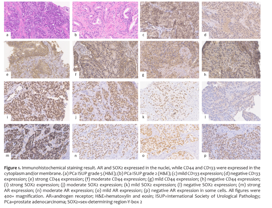
• Immunohistochemistry for CD133, CD44, SOX2, and androgen receptor, scored using H-score.

Key Results

• ISUP grade and PSA level differed significantly between high- and low-volume groups.

• SOX2 expression was significantly associated with metastatic extent.

• The low-volume group showed higher SOX2 expression in the primary tumor.

Discussion & Conclusion

• SOX2 expression in the primary tumor is linked to metastasis extent in castration-naive prostate cancer.

• Different SOX2 levels across sites and stages may reflect the cancer cells’ systemic network.

Related SDGs

SDG 3: Good Health and Well-Being – Advancing understanding of prostate cancer progression

Journal Information

Journal: Medical Journal of Indonesia

DOI: https://doi.org/10.13181/mji.oa.247588

Optimising exercise intensity for gut health: Effect on microbiota composition, barrier integrity and inflammation inmale Wistar rats

Optimising exercise intensity for gut health: Effect on microbiota composition, barrier integrity and inflammation inmale Wistar rats

41

Authors

Nova Sylviana1,2,3 12a92c10 be22 45ce 9934 959ad196a71f       Putri Karisa4 0cae7970 6801 4800 8c04 2a207fd569ca Nur Faizah Romadona5  Imam Megantara2,6

Affiliations

1Physiology Division, Department of Biomedical Sciences, Faculty of Medicine, Universitas Padjadjaran, Bandung, Indonesia 2Physiolgy Molecular Laboratory, Biology Activity Division, Central Laboratory, Universitas Padjadjaran, Sumedang, West Java, Indonesia 3Center of Sport Science, Wellness, and Longevity, Graduate School, Universitas Padjadjaran, Bandung, West Java, Indonesia 4Doctoral Program in Medical Science, Faculty of Medicine, Universitas Padjadjaran, Bandung, Indonesia 5Faculty of Medicine, Universitas Pendidikan Indonesia, Bandung, West Java, Indonesia 6Microbiology Division, Department of Biomedical Sciences, Faculty of Medicine, Universitas Padjadjaran, Bandung, Indonesia

Background

• Exercise influences gut microbiota and intestinal permeability, but the optimal intensity for gut health is unclear.

Methods

• Male Wistar rats were assigned to control, low (10 m/min), moderate (20 m/min), or high (30 m/min) exercise, five times a week for 8 weeks.

• qPCR measured A. muciniphila, F. prausnitzii, and E. coli; ZO-1 (barrier) and IL-6 (inflammation) mRNA were also assessed.

Key Results

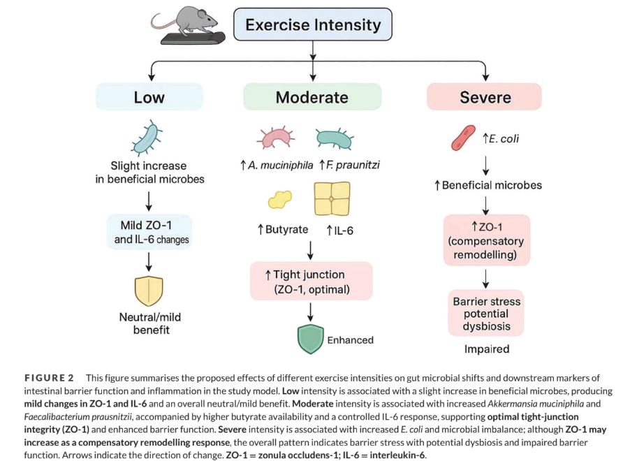
• Moderate intensity increased beneficial A. muciniphila and F. prausnitzii.

• High intensity reduced them and sharply increased E. coli.

• ZO-1 rose at moderate and high intensity; IL-6 also rose, most strongly at high intensity.

Discussion & Conclusion

• Moderate exercise enhanced beneficial gut bacteria and barrier integrity.

• High-intensity exercise promoted E. coli growth and IL-6-mediated inflammation, showing a dose-dependent effect.

Related SDGs

SDG 3: Good Health and Well-Being – Promoting health through optimal physical activity

Journal Information

Journal: Experimental Physiology

DOI: https://doi.org/10.1113/EP092970

Genomic Profile of Interleukin-6 Secreting Ovarian Cancer: Validation of Future Biomarkers and Target of Therapy

Genomic Profile of Interleukin-6 Secreting Ovarian Cancer: Validation of Future Biomarkers and Target of Therapy

40

Authors
Muhammad Ary Zucha1,2, Vincentius Simeon Weo Budhyanto3, Widha Apriani Priadi1, Faizah Dwi Tirtasari4, Moh Nailul Fahmi1, Addin Trirahmanto1, Ardhanu Kusumanto1, Heru Pradjatmo1, Siti Salima b6ec77db d894 4caa a22e dbe620fb7d422, Ali Budi Harsono 6c3b642a 8830 4dbd 9256 16f2555f93152, Gatot Nyarumenteng Adhipurnawan Winarno 79693bd6 af80 429b b63e 80451d84f37e2, Dodi Suardi2, Nadia Yasmine5, Agil Wahyu Wicaksono 73683ead 8bc8 4941 8740 0aca24ba997e6, Laurens David Paulus7, Muhammad Yusuf8, Andi Kurniadi b0b0a042 65fb 49d3 8de0 ade8ddbef6294

Affiliations

1Department of Obstetrics and Gynecology, Division of Oncology Gynecology, Universitas Gadjah Mada – Sardjito Central Hospital, Yogyakarta, Indonesia; 2Department of Obstetrics and Gynecology, Division of Oncology Gynecology Universitas Padjadjaran-Hasan Sadikin Central Hospital, Bandung, West Java, Indonesia; 3Department of Human Genetics, Yayasan Satriabudi Dharma Setia, Tangerang, Banten, Indonesia; 4Department of Pathology Anatomy, Faculty of Medicine, Public Health and Nursing, Universitas Gadjah Mada, Yogyakarta, Indonesia; 5Department of Nutrition, Faculty of Medicine, Public Health and Nursing, Universitas Gadjah Mada, Yogyakarta, Indonesia; 6Department of Biomedical Science, Faculty of Medicine, Universitas Institut Pertanian Bogor, Bogor, West Java, Indonesia; 7Department of Obstetrics and Gynecology, Prof. WZ. Johanes Hospital, Nusa Tenggara Timur, Indonesia; 8Department of Obstetrics and Gynecology, Dharmais Central Hospital, Jakarta, Indonesia

Background

• Inflammation plays a central role in ovarian cancer, and IL-6-secreting tumors may carry altered genetic profiles.

• Studies combining IL-6 measurement with genomic profiling are lacking.

Methods

• 45 women undergoing surgery for ovarian tumors (18 malignant, 19 benign, 8 borderline).

• IL-6 was measured in serum and tumor fluid by ELISA.

• High-IL-6 cancers underwent whole-genome sequencing (ONT).

Key Results

• IL-6 levels were consistent between serum and tumor fluid.

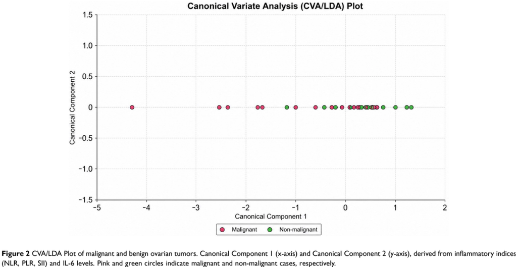
• Malignant tumors secreted significantly more IL-6 than benign ones (p = 0.027).

• IL-6-secreting cancers showed alterations in genes including MTRF1, IL-10, IL-6, APC, PAX8, PTGS2, and PACERR.

Discussion & Conclusion

• Ovarian malignancy is marked by high secretion of pro-inflammatory cytokines.

• Inflammation may disrupt the genetic landscape, offering candidate biomarkers and therapeutic targets for future validation.

Related SDGs

SDG 3: Good Health and Well-Being – Advancing biomarker discovery in ovarian cancer

Journal Information

Journal: International Journal of Women’s Health

DOI: https://doi.org/10.2147/IJWH.S589918

Pin It on Pinterest