
$

PAUS ID

Padjadjaran Authentication System.

$

Portal Mahasiswa

Merupakan portal informasi bagi mahasiwa UNPAD

$

Portal Staff

Merupakan portal informasi bagi staf UNPAD.

$

Email UNPAD

Layanan email berbasis Google App for Education (GAFE) bagi seluruh sivitas akademika Unpad.

UNPAD

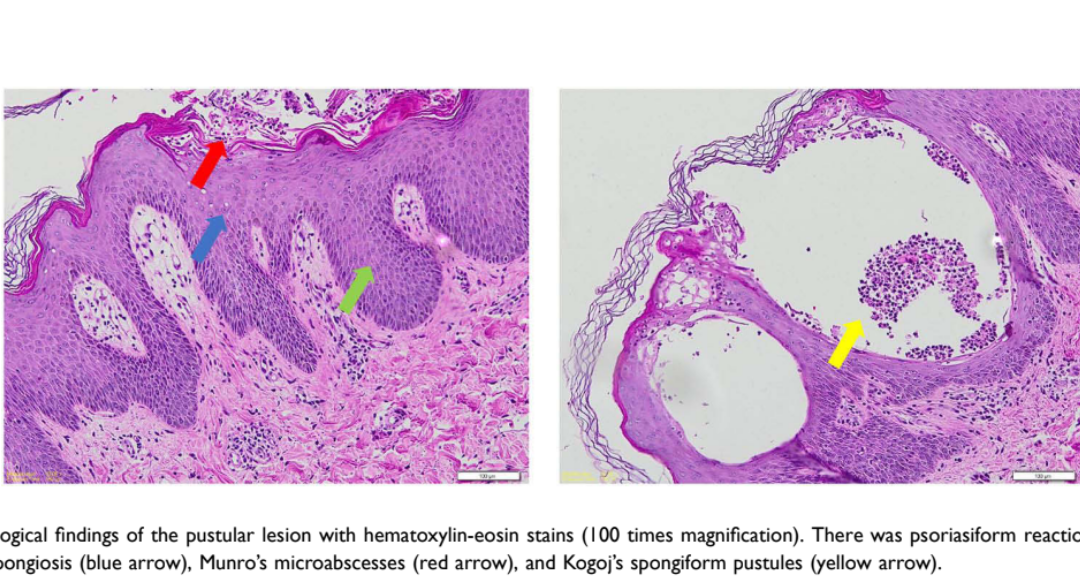
The Uncommon Coexistence of Mid-Borderline Leprosy and Generalized Pustular Psoriasis

The Uncommon Coexistence of Mid-Borderline Leprosy and Generalized Pustular Psoriasis

49

Authors
Hendra Gunawan 1, Risa Miliawati Nurul Hidayah 1, Reiva Farah Dwiyana 1,Trustia Rizqandaru 1, Reti Hindritiani 1, Hermin Aminah Usman 2, Ranisa Larasati 1

Affiliations

1Department of Dermatology and Venereology, Faculty of Medicine, Universitas Padjadjaran – Dr. Hasan Sadikin General Hospital, Bandung, West Java, Indonesia; 2Department of Pathological Anatomy, Faculty of Medicine, Universitas Padjadjaran – Dr. Hasan Sadikin General Hospital, Bandung, West Java, Indonesia

Background

• Leprosy is a chronic disease affecting the skin and nerves, while generalized pustular psoriasis (GPP) is a rare, severe autoimmune skin condition.

• Their coexistence is extremely rare due to very different genetic and immune profiles.

Methods

• Case report of a 28-year-old woman presenting with both mid-borderline leprosy and severe generalized pustular psoriasis.

• Diagnosis combined slit-skin smear, Gram staining, and histopathology of both anesthetic patches and pustular lesions.

Key Results

• Slit-skin smear showed a bacterial index of 1+, and Gram staining of pustules showed no bacteria.

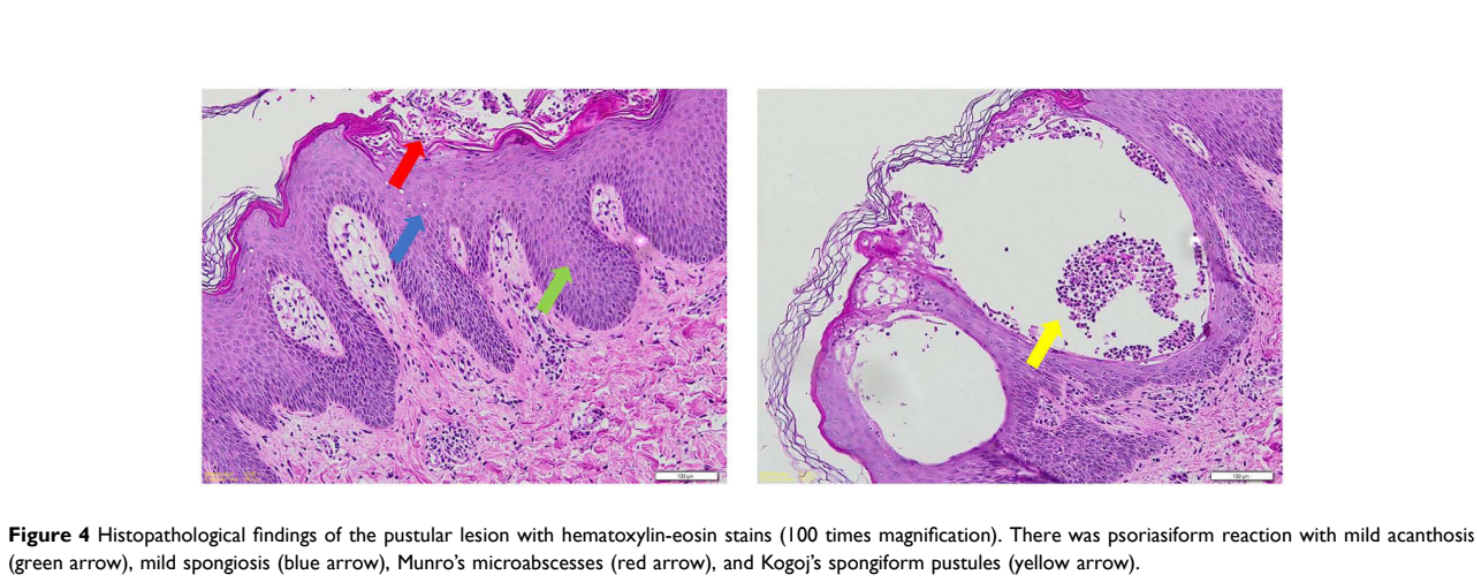
• Histopathology confirmed leprosy granulomas in one set of lesions and features of generalized pustular psoriasis in another.

• She was treated with WHO multidrug therapy for leprosy plus systemic corticosteroids, with marked improvement within 47 days.

Discussion & Conclusion

• This case highlights the importance of recognizing rare coexisting skin conditions with very different underlying mechanisms.

• A comprehensive diagnostic approach and prompt combined management led to a favourable outcome.

Related SDGs

SDG 3: Good Health and Well-Being – Improving diagnosis of complex, overlapping skin diseases

Journal Information

Journal: Clinical Cosmetic and Investigational Dermatology

DOI: https://doi.org/10.2147/CCID.S599655

The Uncommon Coexistence of Mid-Borderline Leprosy and Generalized Pustular Psoriasis

The Uncommon Coexistence of Mid-Borderline Leprosy and Generalized Pustular Psoriasis

48

Authors

Hendra Gunawan1, Risa Miliawati Nurul Hidayah1, Reiva Farah Dwiyana1, Trustia Rizqandaru1, Reti Hindritiani1, Hermin Aminah Usman2, Ranisa Larasati1

Affiliations

1Department of Dermatology and Venereology, Faculty of Medicine, Universitas Padjadjaran – Dr. Hasan Sadikin General Hospital, Bandung, West Java, Indonesia; 2Department of Pathological Anatomy, Faculty of Medicine, Universitas Padjadjaran – Dr. Hasan Sadikin General Hospital, Bandung, West Java, Indonesia

Background

• Leprosy is a chronic disease affecting the skin and nerves, while generalized pustular psoriasis (GPP) is a rare, severe autoimmune skin condition.

• Their coexistence is extremely rare due to very different genetic and immune profiles.

Methods

• Case report of a 28-year-old woman presenting with both mid-borderline leprosy and severe generalized pustular psoriasis.

• Diagnosis combined slit-skin smear, Gram staining, and histopathology of both anesthetic patches and pustular lesions.

Key Results

• Slit-skin smear showed a bacterial index of 1+, and Gram staining of pustules showed no bacteria.

• Histopathology confirmed leprosy granulomas in one set of lesions and features of generalized pustular psoriasis in another.

• She was treated with WHO multidrug therapy for leprosy plus systemic corticosteroids, with marked improvement within 47 days.

Discussion & Conclusion

• This case highlights the importance of recognizing rare coexisting skin conditions with very different underlying mechanisms.

• A comprehensive diagnostic approach and prompt combined management led to a favourable outcome.

Related SDGs

SDG 3: Good Health and Well-Being – Improving diagnosis of complex, overlapping skin diseases

Journal Information

Journal: Clinical Cosmetic and Investigational Dermatology
DOI: https://doi.org/10.2147/CCID.S599655

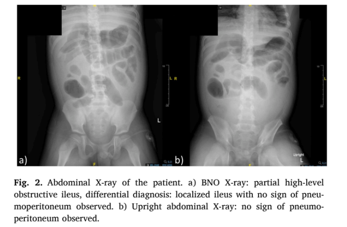
Managing concurrent nephrotic syndrome, spontaneous bacterial peritonitis, and herpes zoster in an immunocompromised child: Diagnostic difficulties and treatment outcomes

Managing concurrent nephrotic syndrome, spontaneous bacterial peritonitis, and herpes zoster in an immunocompromised child: Diagnostic difficulties and treatment outcomes

47

Authors

Riyadi Adrizain a,*69946c28 40eb 44f4 a6b1 ac6e6f16c28d, Ismiana Modjaningrat c, Winyarti c, Fadila Dyah Trie Utami a, Wilson Surya Lesmana d, Vita Indriasari b, Dedi Rachmadi Sjambas a

Affiliations

a Department of Child Health, Faculty of Medicine, Universitas Padjadjaran/Hasan Sadikin General Hospital, Bandung, West Java, Indonesia b Pediatric Surgery Division, Department of Surgery, Faculty of Medicine, Universitas Padjadjaran/Hasan Sadikin General Hospital, Bandung, West Java, Indonesia c Pediatric Resident, Department of Child Health, Faculty of Medicine, Universitas Padjadjaran/Hasan Sadikin General Hospital, Bandung, West Java, Indonesia d Pediatric Surgery Resident, Department of Surgery, Faculty of Medicine, Universitas Padjadjaran/Hasan Sadikin General Hospital, Bandung, West Java, Indonesia

Background

• Nephrotic syndrome (NS) involves heavy protein loss in urine, and poorly controlled disease can lead to serious infectious complications.

• Immunocompromised children with NS face added risk from opportunistic infections.

Methods

• Case report of a 9-year-old boy with a history of nephrotic syndrome since age 7, poorly controlled due to irregular medication use.

• He presented with worsening abdominal pain, fever, and facial oedema, and was managed through hospitalization with imaging, lavage drainage, and antibiotic therapy.

Key Results

• The patient was diagnosed with spontaneous bacterial peritonitis and septic shock related to uncontrolled nephrotic syndrome.

• During hospitalization he developed herpes zoster skin lesions linked to his immunocompromised state.

• He improved with antibiotics and supportive care and was discharged with outpatient follow-up.

Discussion & Conclusion

• This case highlights the importance of early, aggressive intervention in immunocompromised children with nephrotic syndrome facing multiple infectious complications.

• Regular medication adherence is critical to preventing this kind of double infectious burden.

Related SDGs

SDG 3: Good Health and Well-Being – Improving care for children with complex kidney disease

Journal Information

Journal: Idcases

DOI: https://doi.org/10.1016/j.idcr.2026.e02489

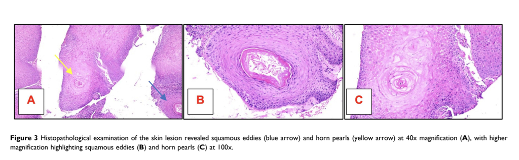
Inverted Follicular Keratosis in Young Adult: A Rare Entity in the Differential Diagnosis of SkinTumors

Inverted Follicular Keratosis in Young Adult: A Rare Entity in the Differential Diagnosis of SkinTumors

46

Authors

Eva Krishna Sutedja c6483897 c2ff 4425 a5d9 4afbcb2b90721, Endang Sutedja 212d7dcd a993 460b a07e 681dc51d05f41, Kartika Ruchiatan1, Yuri Yogya d21197e5 357a 4c34 a822 0684198353ad1, Yogi Faldian 83589aad 9c49 41f5 83fe 4790cddfce891, Oki Suwarsa 325fee8c 8b9c 44b9 afef b9acc9c521f61, Risa Miliawati Nurul Hidayah 7616383e c0f3 49ec 829f ca8f7ba761fa1, Hermin Aminah Usman 1761a496 8cfc 4dcd a713 e23aa2c73dfb2, Erza Rahayu1

Affiliations

1Department of Dermatology and Venereology, Faculty of Medicine, Universitas Padjadjaran – Dr. Hasan Sadikin Hospital, Bandung, West Java, Indonesia; 2Department of Anatomical Pathology, Faculty of Medicine, Universitas Padjadjaran – Dr. Hasan Sadikin General Hospital, Bandung, Indonesia

Background

• Inverted follicular keratosis (IFK) is a rare benign tumor of the hair follicle, usually seen on the head and neck of older adults.

• Its true incidence is likely underreported because it is often misdiagnosed as more common skin lesions.

Methods

• Case report of a 26-year-old man with a solitary, painless, pedunculated scalp lesion that bled intermittently and grew slowly over three years.

• Evaluation included dermoscopy and histopathological examination.

Key Results

• The lesion was initially diagnosed clinically as pyogenic granuloma based on its appearance and bleeding tendency.

• Dermoscopy showed white structureless areas with central keratinous plugs and blood spots.

• Histopathology confirmed inverted follicular keratosis, showing endophytic proliferation and squamous eddies.

Discussion & Conclusion

• IFK can mimic both benign and malignant skin tumors and should remain a differential diagnosis for scalp lesions across all age groups, not just older adults.

• Recognizing its dermoscopic and histopathologic features is critical to avoid misclassification, and histopathology remains essential for a definitive diagnosis.

Related SDGs

SDG 3: Good Health and Well-Being – Improving accurate diagnosis of rare skin tumors

Journal Information

Journal: Clinical Cosmetic and Investigational Dermatology
DOI: https://doi.org/10.2147/CCID.S566801

Efficacy of Vaginal Misoprostol for Labor Inductionin Intrauterine Fetal Death: A Cohort Study from West Java Largest Hospital

Efficacy of Vaginal Misoprostol for Labor Inductionin Intrauterine Fetal Death: A Cohort Study from West Java Largest Hospital

45

Authors

Vania Flowerina *, Dini Hidayat *, Muhammad Alamsyah Aziz *

Affiliations

Obstetrics and Gynaecology Department, Faculty of Medicine Universitas Padjadjaran – Dr. Hasan Sadikin General Hospital, Bandung, Indonesia

Background

• Intrauterine fetal death (IUFD) at 20 weeks or later requires safe and effective labor induction.

• This study assessed the efficacy of vaginal misoprostol.

Methods

• Retrospective cohort at Dr. Hasan Sadikin General Hospital, Bandung (2021–2023).

• 71 women with IUFD at ≥20 weeks who received misoprostol-only induction, compared across gestational age groups (21–27, 28–36, and ≥37 weeks).

• Outcomes: induction-to-contraction interval, induction-to-delivery interval, and cumulative dose.

Key Results

• Median induction-to-contraction time was 12, 6, and 3 hours across the three groups.

• Median induction-to-delivery time was 13, 10, and 6 hours.

• Median cumulative dose fell from 200 µg to 100 µg to 50 µg with advancing gestational age.

Discussion & Conclusion

• Vaginal misoprostol was associated with shorter induction intervals and lower doses at later gestational ages.

• Prospective multicenter studies are needed to confirm optimal regimens and safety.

Related SDGs

SDG 3: Good Health and Well-Being – Improving maternal care in pregnancy loss

Journal Information

Journal: International Journal of Women’s Health

DOI: https://doi.org/10.2147/IJWH.S603527

Pin It on Pinterest