

Authors
Eva Krishna Sutedja 1, Endang Sutedja
1, Kartika Ruchiatan1, Yuri Yogya
1, Yogi Faldian
1, Oki Suwarsa
1, Risa Miliawati Nurul Hidayah
1, Hermin Aminah Usman
2, Erza Rahayu1
Affiliations
1Department of Dermatology and Venereology, Faculty of Medicine, Universitas Padjadjaran – Dr. Hasan Sadikin Hospital, Bandung, West Java, Indonesia; 2Department of Anatomical Pathology, Faculty of Medicine, Universitas Padjadjaran – Dr. Hasan Sadikin General Hospital, Bandung, Indonesia
Background
• Inverted follicular keratosis (IFK) is a rare benign tumor of the hair follicle, usually seen on the head and neck of older adults.
• Its true incidence is likely underreported because it is often misdiagnosed as more common skin lesions.
Methods
• Case report of a 26-year-old man with a solitary, painless, pedunculated scalp lesion that bled intermittently and grew slowly over three years.
• Evaluation included dermoscopy and histopathological examination.
Key Results
• The lesion was initially diagnosed clinically as pyogenic granuloma based on its appearance and bleeding tendency.
• Dermoscopy showed white structureless areas with central keratinous plugs and blood spots.
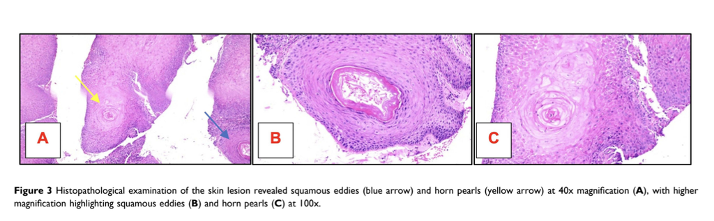
• Histopathology confirmed inverted follicular keratosis, showing endophytic proliferation and squamous eddies.
Discussion & Conclusion
• IFK can mimic both benign and malignant skin tumors and should remain a differential diagnosis for scalp lesions across all age groups, not just older adults.
• Recognizing its dermoscopic and histopathologic features is critical to avoid misclassification, and histopathology remains essential for a definitive diagnosis.
Related SDGs
SDG 3: Good Health and Well-Being – Improving accurate diagnosis of rare skin tumors
Journal Information
Journal: Clinical Cosmetic and Investigational Dermatology




0 Comments